Dental X-Rays in Emergencies: Why They Matter and What They Show
Dental X-rays are essential for accurate emergency diagnosis. American Urgent Dental in Alexandria, VA and Greenbelt, MD explains what emergency X-rays show and why they're safe.
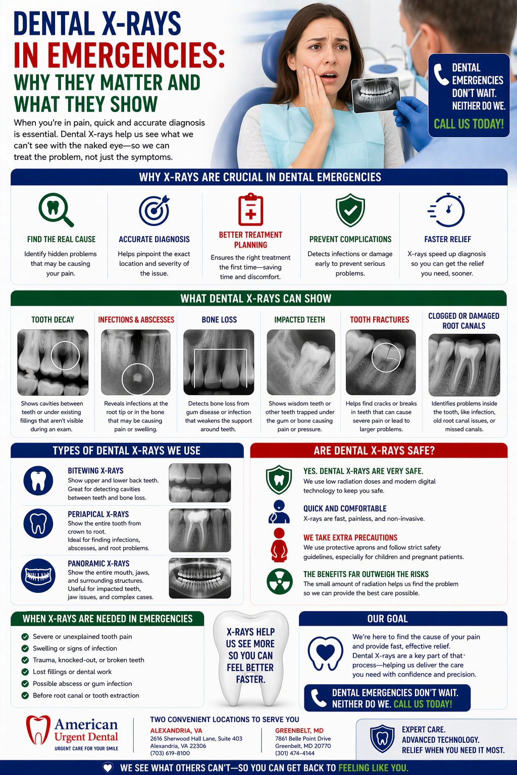
Emergency Dental X-Rays: Seeing What Symptoms Can't Tell You
When you arrive at American Urgent Dental with a dental emergency, one of the first things we'll do — even before treatment begins — is take digital X-rays. Some patients wonder why this is necessary, particularly if they're in significant pain and just want it fixed. Others have concerns about radiation. And some wonder what the X-rays actually show that looking in the mouth doesn't.
This guide explains why diagnostic X-rays are not a bureaucratic step or a revenue-generating add-on — they are essential clinical information that directly determines the right treatment for your emergency, and without them, we would be making treatment decisions in the dark.
What Dental X-Rays Show That Visual Examination Cannot
The Inside of the Tooth
The tooth's exterior — enamel and visible crown structure — is only a small part of the clinical picture. Most dental emergencies involve pathology that is entirely inside the tooth or at the root tip, beneath the bone: dying pulp tissue, infected root canal space, periapical abscesses, root fractures, and the extent of internal decay. None of these can be visualized by looking in the mouth. X-rays are the only way to see them.
The Root and Root Tip
The root of a tooth — which constitutes approximately 60% of the tooth's total length — is entirely within the bone and gum tissue. Root anatomy determines how many canals a root canal must address. Root position determines extraction technique. Root proximity to the sinus floor or inferior alveolar nerve determines surgical risk. Root fractures (vertical root fractures) are often only apparent on X-ray. All of this is invisible without imaging.
The Surrounding Bone
The bone supporting a tooth is the foundation of its long-term survival. Bone changes on X-ray — a dark shadow (radiolucency) at the root tip indicating an abscess, widening of the periodontal ligament space indicating infection, crestal bone loss indicating gum disease, or a fracture through the alveolar bone — directly inform diagnosis and treatment planning. A tooth that appears externally intact may have already lost significant bone support, making it non-restorable.
Adjacent Structures
Emergency treatment decisions must account for proximity to anatomical structures that must not be damaged:
- The inferior alveolar nerve (running through the lower jaw) — risk of permanent paresthesia (numbness) if a lower molar extraction inadvertently contacts it
- The maxillary sinus (above upper molar roots) — perforation into the sinus during upper molar extraction requires specific management
- Adjacent tooth roots — excessive force toward an adjacent tooth can damage its root
- The mental foramen (where the mental nerve exits the lower jaw) — in the vicinity of lower premolar extractions
These relationships can only be assessed with X-rays. A dentist who skips imaging before an extraction is practicing below the standard of care.
Types of X-Rays Used in Emergency Dentistry
Periapical X-Ray
The most important X-ray for emergency dentistry. A periapical X-ray shows the entire tooth from crown to root tip and the surrounding bone. This is the primary image for diagnosing abscesses, root fractures, periapical pathology, pulp status, and root anatomy. We take periapical X-rays of every tooth we treat in an emergency context.
Panoramic X-Ray (OPG)
A single image that shows all the teeth, both jaws, the sinuses, and the temporomandibular joints. This is the most useful image when the source of pain is unclear, when we need to evaluate the whole dentition, when evaluating jaw fractures or facial trauma, or for planning wisdom tooth assessment. Not always needed for simple emergencies, but invaluable for complex presentations.
Bitewing X-Rays
Show the crowns of upper and lower teeth together, primarily useful for detecting interproximal (between-tooth) decay and assessing the height of the alveolar bone. Less commonly needed in pure emergency situations but sometimes used when decay assessment is part of the emergency evaluation.
Radiation Safety: What the Actual Numbers Mean
Radiation concerns are legitimate and deserve honest answers rather than dismissal. Here are the facts:
- A single digital periapical dental X-ray delivers approximately 0.005 millisieverts (mSv) of radiation
- A standard chest X-ray delivers approximately 0.1 mSv — 20 times the dose of a single dental X-ray
- The annual radiation dose from natural background sources (cosmic radiation, radon, etc.) in the United States is approximately 3 mSv — equivalent to 600 dental X-rays
- A full set of dental X-rays (18 films) delivers approximately 0.1 mSv — equivalent to a single chest X-ray
- Modern digital X-ray systems use 50–80% less radiation than older film-based systems
The radiation from emergency dental X-rays is, in plain terms, minimal. The clinical benefit of accurate diagnosis vastly outweighs any theoretical radiation risk from the small doses involved.
X-Rays During Pregnancy: The Safe Answer
A concern we frequently hear from pregnant patients: 'I don't want X-rays because I'm pregnant.' This is an understandable concern, and the honest answer is: dental X-rays during pregnancy are safe when performed with appropriate shielding. With a thyroid collar over the thyroid gland and a lead apron over the abdomen, fetal radiation exposure from dental X-rays is essentially zero. The American Dental Association and the American Congress of Obstetricians and Gynecologists both explicitly support dental X-rays in pregnancy when clinically indicated — particularly for dental emergencies where the information guides treatment decisions affecting maternal health.
What Happens When We Skip X-Rays
The consequences of treating dental emergencies without X-rays range from suboptimal to dangerous:
- A tooth extracted without X-ray evaluation of root anatomy breaks during extraction because the root morphology was unknown
- A root canal attempted without X-ray misses a hidden fourth canal, leaving infected tissue that causes recurrent abscess
- An antibiotic prescribed for what 'looks like' an abscess turns out to be a jaw fracture requiring surgical management
- An extraction performed without checking for proximity to the inferior alveolar nerve causes permanent lower lip and chin numbness
We take X-rays because the information they provide makes us better dentists and protects you as a patient. They are part of our standard of care for every emergency patient, and they are part of what makes emergency care at American Urgent Dental genuinely excellent. Call us: Alexandria 703-214-9143 | Greenbelt 240-241-0342.
Get Same-Day Emergency Dental Care
American Urgent Dental — two convenient locations serving Northern Virginia and the Greater DC Metro area.
Alexandria, VA: 2616 Sherwood Hall Lane Ste 403, Alexandria, VA 22306 | 703-214-9143
Greenbelt, MD: 7861 Belle Point Drive, Greenbelt, MD 20770 | 240-241-0342
📧 contact@americanurgentdental.com | 🌐 www.americanurgentdental.com
Don’t wait out the pain.
Same-day emergency care in Alexandria, VA & Greenbelt, MD — open weekends.